A UVU undergraduate has built DIFFRAX, a laser-and-AI diagnostic device that detects bacterial infection in knee implants without surgery. Presented at the 2026 Utah NASA Space Grant Consortium Symposium, the tool achieves 99.2 percent accuracy at a parts cost under $400.
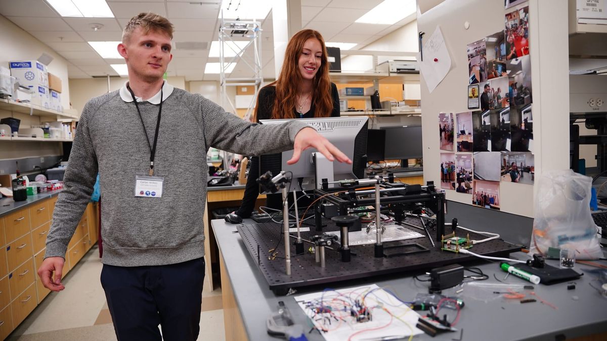
Carter Wilkes demonstrates his DIFFRAX device in the CIBEAM lab at Utah Valley University alongside Adelle Webb, Lead Coordinator at UVU's Kahlert Applied AI Institute (AAII). The machine — built from repurposed 3D printer parts and off-the-shelf components for under $400 — uses a red laser and a custom-trained neural network to detect bacterial infection in knee implants without surgery. Photo: Greg Tullis - Kahlert Applied AI Institute Project Team
Carter Wilkes demonstrates his DIFFRAX device in the CIBEAM lab at Utah Valley University alongside Adelle Webb, Lead Coordinator at UVU's Kahlert Applied AI Institute (AAII). The machine — built from repurposed 3D printer parts and off-the-shelf components for under $400 — uses a red laser and a custom-trained neural network to detect bacterial infection in knee implants without surgery. Photo: Greg Tullis - Kahlert Applied AI Institute Project Team
A UVU student's AI-powered device could end a painful — and costly — diagnostic procedure for joint replacement patients.
Orem, Utah — May 6, 2026
The morning before the symposium opened, Kim Olson got a phone call. A student who was scheduled to present his research that day wouldn't be coming. "I just got a job at SpaceX," he told her, "and I'm leaving on Sunday." Olson's response was immediate: you can miss it. The student — a BYU graduate — pre-recorded his presentation and was gone by the next day.
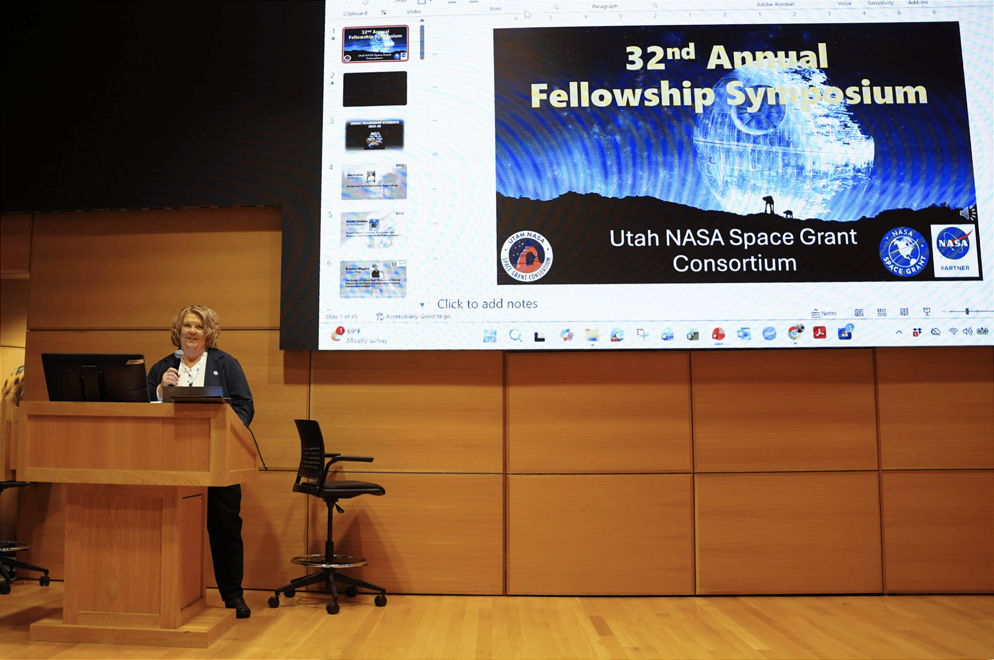
Kim Olson, Associate Director of the Utah NASA Space Grant Consortium, addresses attendees at the 32nd Annual Fellowship Symposium at Utah Valley University on May 4, 2026. Behind her, the event's Star Wars-themed title slide reflects the occasion — the symposium fell on May the Fourth. Olson has helped lead the consortium since its early days, when the fellowship brought together just a handful of graduate students from three Utah universities. This year's event drew researchers from institutions across the state. Photo: Greg Tullis, Kahlert AAII Team
For Olson, Associate Director of the Utah NASA Space Grant Consortium, the call was not a disruption. It was the point. "It's not very much money," she says of the fellowships her program distributes — graduate students receive between $15,000 and $25,000, undergraduates sometimes as little as $1,000. "But it moves them into a place where they can actually do something."
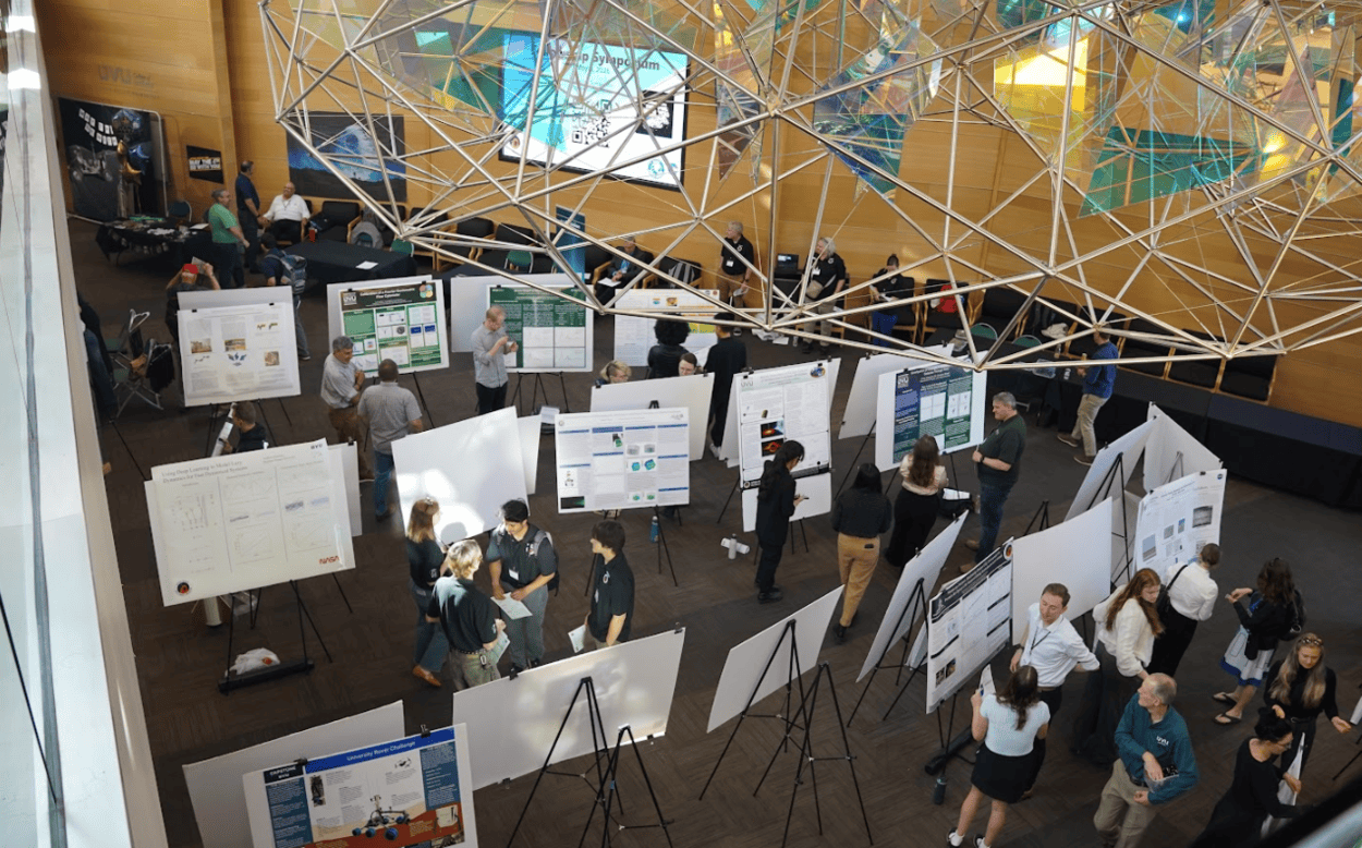
At the 32nd Annual Utah NASA Space Grant Consortium Fellowship Symposium, held May 4, 2026, at Utah Valley University in Orem, the evidence of what that nudge can produce was on display across two floors of the Science Building. Students from universities across Utah — BYU, Utah State, the University of Utah, Snow College, Salt Lake Community College, Utah Tech, and others — presented original research spanning aerospace, robotics, biomedical innovation, and astronomy. One of them showed up with a device that may one day replace a surgical procedure. He built it for under $400.
Researchers, faculty, and industry representatives move through the poster session at the 32nd Annual Utah NASA Space Grant Consortium Fellowship Symposium, held May 4, 2026, in the atrium of Utah Valley University's Science Building in Orem, Utah. Students from universities across Utah presented original research spanning biomedical innovation, aerospace engineering, robotics, and astronomy. Photo: Greg Tullis, Kahlert AAII Team
The Problem No One Wanted to Talk About
When someone receives a knee implant, they begin a quiet waiting game. Infection is a persistent risk — not dramatically common, but catastrophic when it arrives. And for years, the only reliable way to know whether bacterial biofilm had taken hold on the implant was to take an invasive sample from an already sensitive injury. If infection was found, the fix was straightforward: antibiotics. But confirming the presence of infection required surgery. The aggressive procedure was the diagnosis.
Carter Wilkes, a Physics undergraduate at Utah Valley University, is working to make that invasive step unnecessary. Working in UVU's Center for Imaging and Biophotonics Experiments Advancing Medicine — known as CIBEAM — under faculty advisor Dr. Vern Hart, Wilkes and his team have built a device called DIFFRAX (Diffraction Imaging for Film Formation and Reflective Anomaly Extraction). It uses a red laser and a custom-trained neural network to detect bacterial infection through the skin, without cutting or scraping, and in about five minutes.
DIFFRAX isn't the lab's first attempt to reimagine how AI and imaging can detect what was previously invisible. Wilkes has previously worked on RUTID, a rapid UTI detection device, as part of CIBEAM's broader focus on using optics and imaging to make diagnostics faster and less invasive. The knee implant project represents the lab's most clinically urgent application yet.
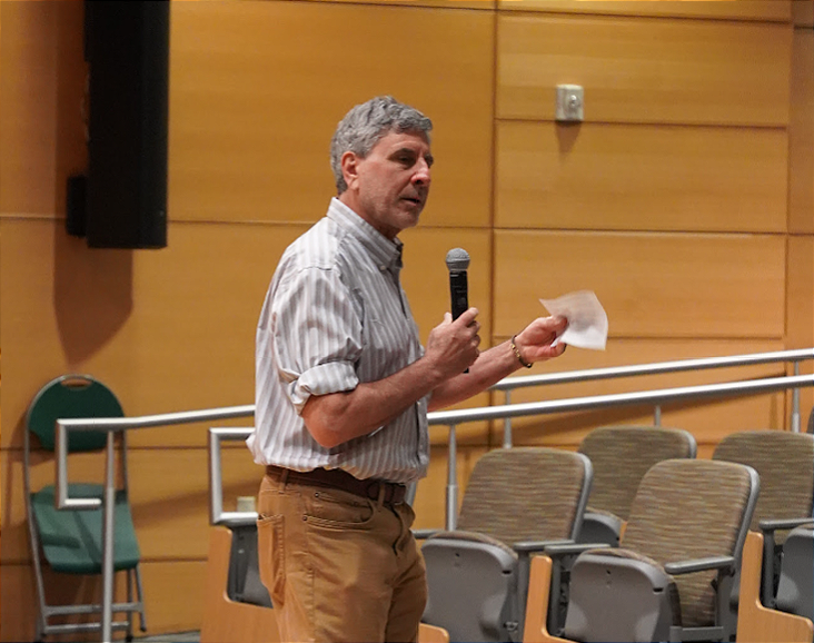
Dr. Daniel Horns, Dean of the College of Science at Utah Valley University, addresses students and faculty at the opening of the 32nd Annual Utah NASA Space Grant Consortium Fellowship Symposium, held May 4, 2026, at UVU's Science Building in Orem. Horns encouraged student presenters to look beyond the day's résumé value. Photo: Greg Tullis - Kahlert AAII Team
Opening the morning's proceedings, Dr. Daniel Horns, Dean of UVU's College of Science, reminded students that the stakes of the day were higher than a line on a résumé. "The fact that you're doing the work that got you here to present a poster or an oral talk means that you're doing research beyond the classroom," he said. "You're learning skills and knowledge that can't be gained in the classroom."
Wilkes is doing exactly that through his novel scanning technology.
Built From Parts, Built for Patients
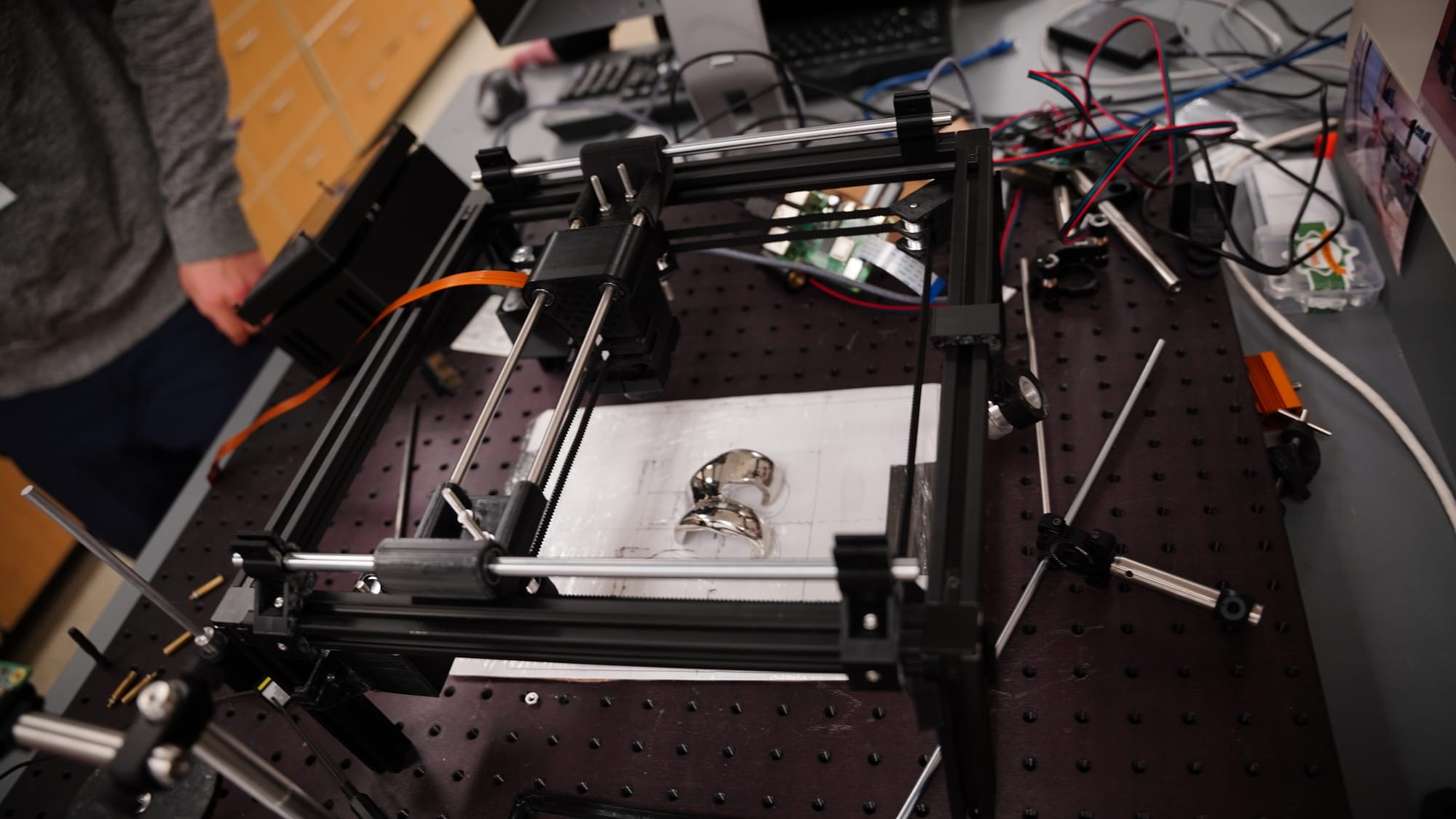
The DIFFRAX scanner doesn't look like medical equipment. Resting on a vibration-dampening optical table in UVU's CIBEAM lab, it resembles what it partly is: a heavily modified 3D printer. The open black aluminum frame, the dual steel rod rails, the belt-and-carriage movement system — the machine's skeleton is unmistakably borrowed from consumer desktop fabrication technology. But where a 3D printer deposits material, DIFFRAX fires a 635nm red diode laser across a scan bed, methodically mapping the surface beneath it point by point.
The DIFFRAX scanner positions a chrome knee implant at the center of its scan bed inside UVU's CIBEAM lab. The device's open aluminum frame and belt-driven rail system were adapted from a 3D printer. A Raspberry Pi 4 (visible, upper center) processes the laser diffraction data in real time. Total cost of parts: under $400. Photo: Greg Tullis - Kahlert AAII Team
At the center of the scan bed, taped carefully to a sheet of paper with hand-drawn alignment markings, sits a chrome knee implant. It is the machine's only patient — for now. The laser travels down, strikes the implant's reflective surface, and bounces back to a CMOS beam profiler mounted above. An orange ribbon cable carries that signal to the device's brain: a Raspberry Pi 4, the same $99 computer used in school science projects and hobbyist electronics kits worldwide. Stitching code assembles each reflected data point into a complete diffraction map of the implant surface.
The entire bill of parts totals less than $400.
In the lab, Wilkes walked through the process with the ease of someone who has operated it many times. The camera mounted at the bottom of the carriage detects photon scattering patterns as the laser fires at the implant below. Two stepper motors drive the carriage across its x and y axes, rastering back and forth in a grid, "literally just like a 3D printer," Wilkes explained. The system needs only 136 images to generate a result with a reliable confidence interval. When the scan is complete, the device displays its findings directly on screen, telling the operator — in Wilkes's words — "with what accuracy confidence interval you have a biofilm." For conferences and eventual clinical use, the entire system runs portably: connect it, press Scan, and wait.
How DIFFRAX Works: Light, Reflection, and a Trained Eye
The physics behind DIFFRAX begins with a property of red light that Wilkes describes with characteristic directness: "You can actually shoot a red laser into skin, and it can get about eight millimeters in before it completely diffuses." For a knee implant positioned just beneath the skin's surface, that penetration depth is precisely what's needed. The laser reaches the implant and reflects back — carrying with it information about what it passed through.
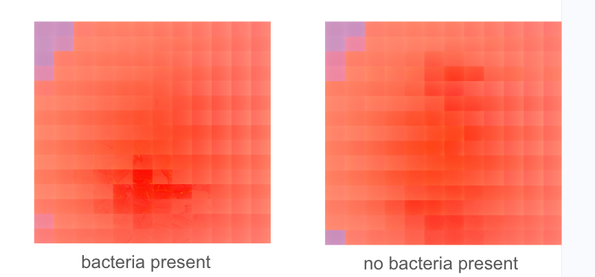
That reflected signal becomes a diffraction map, a pixelated grid image recording how light scattered across the implant surface. To the untrained eye, a clean map and an infected one look nearly identical: abstract arrangements of light and dark squares with no obvious pattern. That invisibility is precisely the problem DIFFRAX was built to solve.
"We have trained a neural network to see the minute differences between a no-bacteria sample and a bacteria sample," Wilkes explained. The system has been trained on thousands of images, learning to detect patterns invisible to human perception — subtle variations in how light scatters and reflects when biofilm is present. In the infected diffraction maps, a dark cross-shaped anomaly emerges at the center, absent in clean samples. A human analyst would likely miss it. The neural network does not. "We wouldn't be able to use this without AI," Wilkes said plainly.
Two diffraction maps produced by the DIFFRAX device. The right image shows a clean implant surface with no bacterial presence. The left image shows the same type of implant with bacterial biofilm — the dark cross-shaped formation at lower center is the anomaly the system's neural network has been trained to identify. To the human eye, the two images appear nearly identical. Image: Carter Wilkes
The results so far are striking. In testing, the system has achieved 99.2 percent accuracy in distinguishing infected from uninfected samples. The patient experience, should the device reach clinical deployment, would be a five-minute scan — non-invasive, repeatable, and schedulable as a routine follow-up. Wilkes envisions patients returning "once a week for three months" after surgery, catching problems early rather than waiting for symptoms to force a more drastic response.
What Early Detection Actually Means
The financial stakes of the problem Wilkes is addressing are not abstract. When bacterial infection in a joint implant goes undetected until it becomes symptomatic, the result is almost always additional surgery. "Those revision surgeries can cost anywhere from $50,000 to $100,000," Wilkes noted. A five-minute weekly scan from a modest machine built from $400 in parts, by comparison, is a rounding error — and that contrast is not lost on the team.
But the more significant cost is the one that doesn't appear on a bill. Patients who develop serious post-implant infections often face months of additional treatment, prolonged immobility, and in severe cases, implant removal. For patients who have already endured major surgery, early detection through a noninvasive tool like DIFFRAX wouldn't just save money—it could reshape the path to recovery.
The project is labeled "Bench to Bedside" on the DIFFRAX poster, a deliberate framing that signals the team's intent to move the research beyond the lab. Biofilms, the poster notes, are responsible for over 60 percent of hospital-acquired infections, contributing to hundreds of thousands of implant-related infections annually in the U.S. The CIBEAM team, which includes more than a dozen student researchers alongside Dr. Hart, is building the evidence base that a future clinical or regulatory pathway would require.
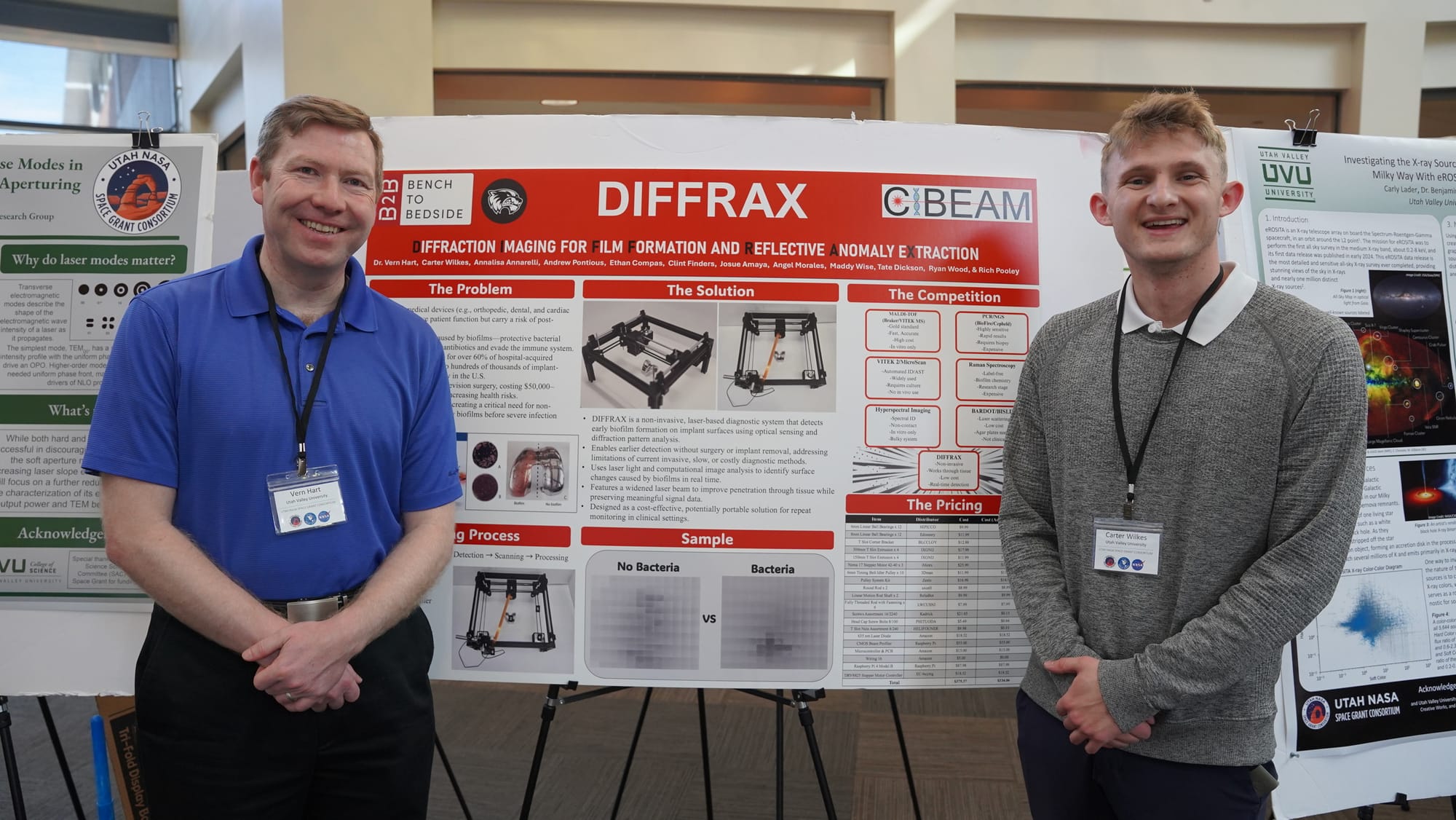
Carter Wilkes (right) and faculty advisor Dr. Vern Hart stand before the DIFFRAX research poster at the 32nd Annual Utah NASA Space Grant Consortium Fellowship Symposium, held May 4, 2026, at Utah Valley University. Wilkes built the laser-based diagnostic device for under $400 in parts. In testing, it has identified bacterial infection in knee implants with 99.2 percent accuracy — without surgery. Photo: Greg Tullis, Kahlert AAII Team
Across the Symposium: Technology at the Edge of What's Known
The DIFFRAX project was the most clinically immediate of the day's presentations, but it was not alone in using computational tools to solve problems that previous methods couldn't reach.
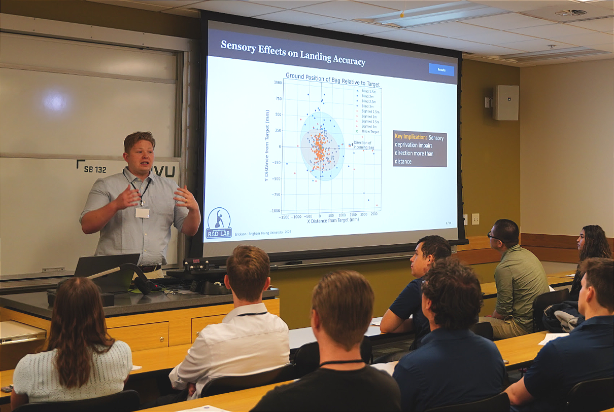
Brigham Young University student Austin Erickson presented research on a deceptively simple task: two people throwing an object together. His work, conducted in BYU's RAD Lab under Dr. Marc Killpack, focused on how human coordination actually emerges in this scenario — and what it means for robotics. "Throwing is a really interesting task where the whole performance is determined by a single release event," Erickson explained. His data, drawn from both sighted and blindfolded throwing trials, revealed a finding that surprised even seasoned observers: sensory deprivation impairs directional accuracy more than distance. Blindfolded throwers still landed close to the target — they just got the angle wrong. "Coordination emerges through a sensing of motion, rather than synchronization and release," Erickson concluded. The implication for robotic systems is significant: rather than programming exact movements, future robots could be designed to respond dynamically to a partner's motion, reading it in real time the way humans instinctively do — even without full sensory information.
Austin Erickson of Brigham Young University presents findings from his dyadic throwing research during Session 1 of the 32nd Annual Utah NASA Space Grant Consortium Fellowship Symposium, held May 4, 2026, at Utah Valley University. His scatter plot data, comparing throw accuracy between sighted and blindfolded participants, revealed that sensory deprivation impairs directional accuracy more than distance, a finding with direct implications for how robots might be designed to sense and respond to a partner's motion in real time. Photo: Greg Tullis, Kahlert AAII Team
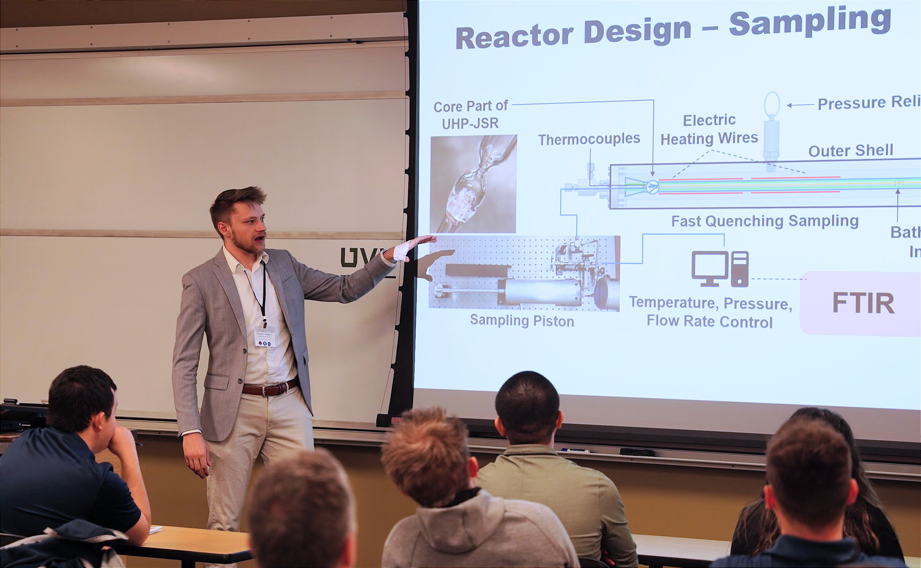
Braxton Wiggins, a graduate student at Utah State University, presented work on ultra-high-pressure combustion systems — specifically, designing a jet-stirred reactor capable of testing fuels at conditions that current computational models cannot reliably simulate. The reactor itself is a precisely engineered system: thermocouples and electric heating wires regulate conditions inside a pressurized outer shell, while a fast-quenching sampling piston captures reaction products mid-combustion for analysis by an FTIR spectrometer. The fuels under study include hydrogen, methane, and ammonia. They are candidates relevant to next-generation propulsion systems. "Once you get to above the supercritical pressure points of these fuels, the computations get very complicated and intensive and don't exactly know if they're right," Wiggins explained. His physical testing apparatus exists to validate and correct those models using chemical kinetic software including Cantera and ANSYS Chemkin-Pro, with direct implications for the efficiency of rocket engines operating at extreme conditions.
Braxton Wiggins, a graduate student at Utah State University, presents the design of his Ultra-High-Pressure Jet-Stirred Reactor during the 32nd Annual Utah NASA Space Grant Consortium Fellowship Symposium at Utah Valley University. His reactor is designed to test fuels at supercritical pressures — conditions where current computational models become unreliable — with the goal of improving the accuracy of rocket engine efficiency calculations. Photo: Greg Tullis, Kahlert AAII Team
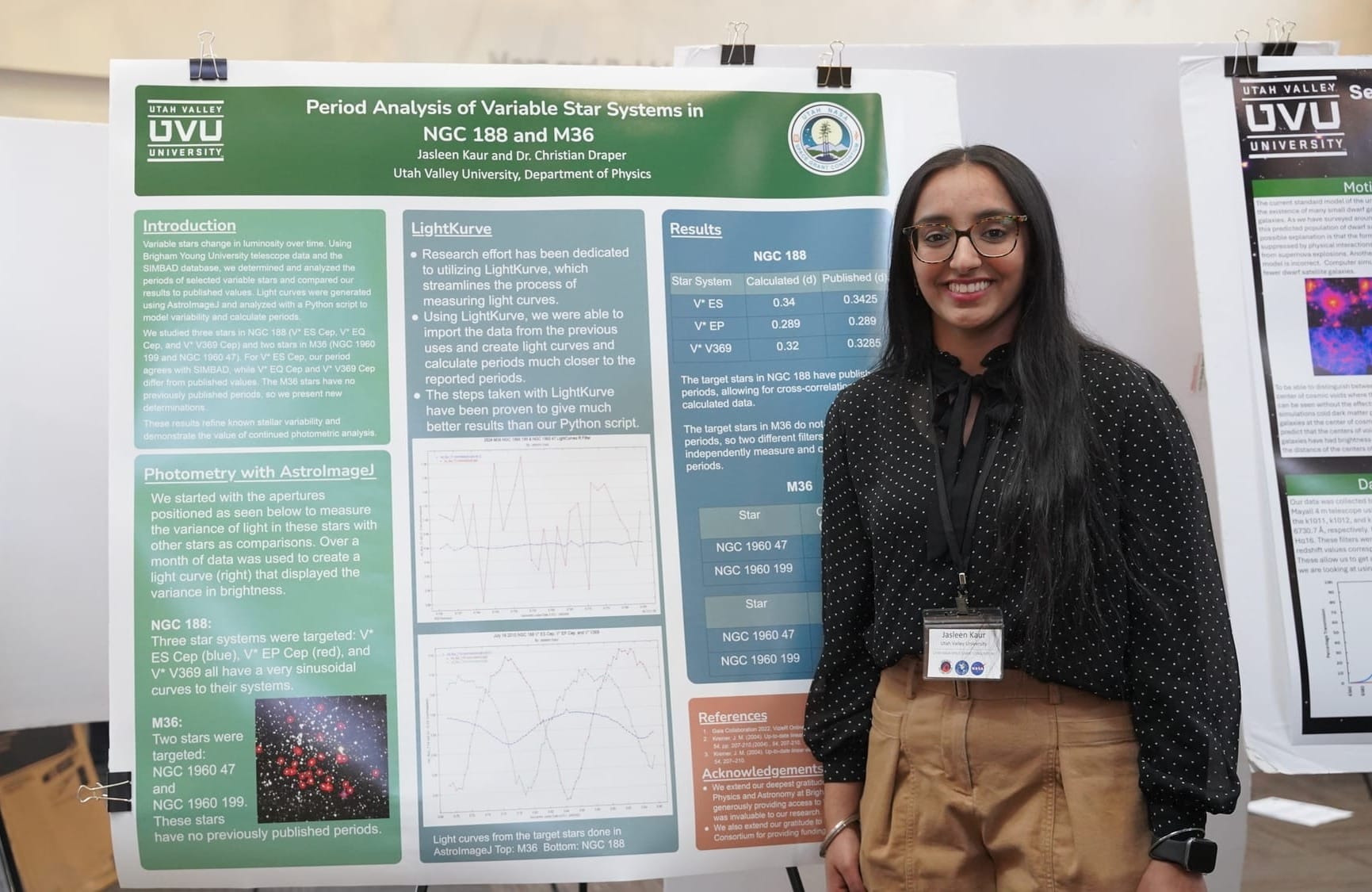
UVU student Jasleen Kaur brought a different scale of inquiry to the symposium's poster session — not the interior of a knee, but the interior of a galaxy. Working under Dr. Christian Draper in UVU's Department of Physics, Kaur is analyzing variable stars in two open star clusters, NGC 188 and M36, using telescope data from BYU's West Mountain Observatory processed through Python-based photometry tools. For M36, no published rotational period exists for either of the two stars she is studying; Kaur's calculations are original data. For NGC 188, where the last published measurements date to 2003, her work confirms whether those stars still behave as expected, a quieter but no less necessary contribution. The broader significance lies in what astronomers call standard candles: variable stars whose predictable behavior makes them reliable reference points for measuring distances across space. If those reference points drift and the data isn't updated, Kaur explained, everything calculated from them has to be revised, including galaxy-to-galaxy distance measurements built on decades of prior work.
Jasleen Kaur, a Physics student at Utah Valley University, presents her period analysis of variable stars in open clusters NGC 188 and M36 at the symposium poster session. Her work under Dr. Christian Draper includes the first calculated rotational periods for two stars in M36, measurements with no prior published reference. Photo: Greg Tullis, Kahlert AAII Team
Research Beyond the Classroom
What connected the day's most compelling presentations was not a shared discipline but a shared method: identify the point at which existing tools fail, then build something that works past that limit. For Wilkes, the limit was human perception — no eye, trained or untrained, can see bacterial biofilm through skin, nonetheless detect it with consistent accuracy using a laser combined with AI-enhanced images. For Erickson, it was the assumption that coordination requires communication. For Wiggins, it was the gap between what computational models predict and what actually happens at extreme pressure. For Kaur, it outdated data that could potentially affect the galaxy as we understand it.
In each case, the researchers didn't accept the limit as a boundary. They built instruments — physical, computational, or both — to see past it. In Wilkes's case, that instrument started as a broken 3D printer and a $15 computer, and became a device that may one day spare patients from surgery they never needed.
That disposition, Dr. Horns suggested in his opening remarks, is what distinguishes research from coursework. "You're learning skills and knowledge that can't be gained in the classroom," he told the assembled students. At a symposium where an undergraduate built a potential medical diagnostic tool from salvaged parts and an AI trained to see what human eyes cannot, the point was difficult to argue with.